Removal of skin lesions
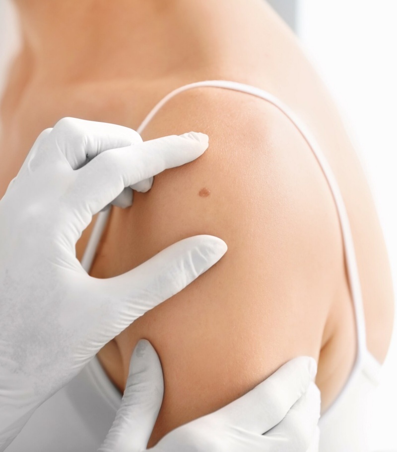
Removal of skin lesions is probably the most common procedure performed by a surgeon in an outpatient setting. Each of us is different, so in daily practice meets various clinical situations. we have the instruments for the possibility of an individual approach to each patientin Cryocentre. The choice of method depends on the nature of the change, its size and location. Before you visit a surgeon, we recommend visit to dermatologist, during which the dermatologist after collecting a detailed history and after evaluation of skin lesions qualify them to be removed in a way that in his opinion is the best, and expected a cosmetic effect will be in line with the expectations of the patient.
Removing CO2 laser skin lesions
The treatment consists of the emission of light energy at a wavelength of 10600 nm. on the surface of the lesion. Operating the laser beam is about 0.1 mm, and the thermal shock of the surrounding tissue is small, which enables very precise operation in the surgical field. We can remove the changes on the surface of the skin with the laser such as:
• Pigmented lesions and other signs
• fibroid tumors, nodules
• Hemangiomas ruby
• cutaneous warts (known. “Warts”)
• Genital warts
• Foot Calluses
• Other skin changes
The procedure is performed under local anesthesia, takes a few to several minutes, it is painless, bloodless and non-contact, which means it does not carry the risk of infection. After surgery the operated place coincides with scabs, which drops evidencing in about 10-14 days a gentle, pink skin. Young skin takes on the color of the surrounding skin over time. The wound heals relatively quickly without leaving a scar or leaving a small, shallow recess with accompanying autumnal at a treatment site (it depends on the size and type of removed change, and the skin phototype).
The removal of skin lesions by cutting
Another way to remove the lesion is a surgical resection, which involves removing a spindle skin with the change while maintaining approximately 1mm margin of healthy tissue. To obtain the best cosmetic result, the cutting lines run along the so-called. Langer’s lines. The resulting loss of tissue is provided with the help of sutures removed approx. 5 – 14 later (depending on the area of the body and the healing process). The resected piece of tissue can be sent to histopathological verification (examination under a microscope), which will assess the nature of the changes, as well as completeness and margins cut. This will allow an assessment of the grade of the affected tissue and can affect any further therapeutic process. The biggest advantage of this type of surgery is obtaining undamaged material for histopathological examination, while the biggest disadvantage is the effect of beauty in the form of scar, which always remains after the intervention.
This method is particularly suitable for:
• skin changes that raise suspicion for malignancy and absolutely require verification of histopathology (such evaluation may be done during dermoscopy prior to surgery)
• lesions that are under the surface of the skin like. Lipoma and atheroma
After surgery
The proper care of the operated area is a very important issue. Failure to follow the doctor’s instructions may affect the cosmetic effect. Avoid damage and mechanical irritation and prolonged soaking the treated surface. Do not apply cosmetics or medicines that may harm or irritate the cells of the newlyepidermis, covering the wound. Exposure of the treated skin to sunlight should be avoid for a period of 2 to 6 months. In the case of the removal of major changes in order to achieve the best results we recommend the use of cosmetic adhesive bandages.


